OBSTETRIC ULTRASOUND

WHAT IS OBSTETRIC ULTRASOUND ?
It is a safe, painless and noninvasive examination that allows the fetus to be visualized on a monitor through the use of a probe, with high-frequency sound waves, resting on the mother's abdomen. When the waves, sent by the probe reach the fetus, they send back other echoes, which are transformed into images on the ultrasound monitor, so that the fetus can be seen in detail.
IS IT HARMLESS TO THE FETUS?
Ultrasound has been used in obstetric practice for more than 30 years and no harmful effects on the fetus have been reported even in the long term. Therefore, with the procedures adopted today, the diagnostic use of ultrasound is considered risk-free.
WHAT IS IT FOR?
At least three obstetric ultrasound examinations should be performed during a physiological pregnancy, in the first trimester (6 weeks to 13 weeks, usually between 11 weeks and 13 weeks), second trimester (19-22 weeks), and third trimester (30-34 weeks), respectively. However, the examination may be repeated during the course of the pregnancy outside of these periods in support of the obstetrical examination or as directed by the physician.
I ULTRASOUND (I TRIMESTER, DATING)
When performing the first-trimester obstetrical ultrasound, generally from weeks 6 to 11 (13), the gynecologist verifies the presence of pregnancy within the uterus, thus providing the correct position of the gestational chamber. She determines the number of embryos, determining whether it is a single or twin pregnancy. It measures the length of the embryo (vertex-sacral length - CRL -) and whether this corresponds to the presumed time of pregnancy, thus indicating the presumed date of delivery. Finally, it confirms the viability of the embryo by viewing cardiac activity.
II ULTRASOUND (II TRIMESTER, MORPHOLOGICAL)
Morphologic ultrasound is performed between 20 and 22 weeks of pregnancy. While conducting the morphological echo, the gynecologist is able to make a complete check of the morphology of the fetus. It checks the fetus' internal organs (head, chest, abdomen, heart, spine, liver, kidneys, bladder, upper and lower limbs), the position of the placenta, and the amount of amniotic fluid. This is the most important examination in pregnancy because of the possibility of assessing possible fetal malformations (central nervous system, neural tube, spine, heart, large vessels, digestive system, urinary system, abdominal wall and limbs). It is believed that a Level I ultrasound examination can detect 30% to 70% of all malformations. It may happen that certain fetal abnormalities escape ultrasound examination. As the possibility of detecting an abnormality depends on its extent, the amount of amniotic fluid, the thickness of the maternal abdominal wall, and finally the position assumed by the fetus inside the uterus during the examination. When faced with a fetus with an "anterior back," ultrasound investigation of ventral anatomical structures may be incomplete. Therefore, further ultrasound evaluation may be required later, to be agreed upon at the time of the examination. Sometimes if the position of the fetus allows, the gynecologist may also reveal the sex of the baby to the parents-to-be.
III ULTRASOUND (III TRIMESTER, GROWTH)
In the third trimester from (28) 30th to (32) 34th weeks the third ultrasound, called "growth" ultrasound, is performed, in which the auxological parameters of the fetus (the measurements of the head - BPD and CC -, abdomen - CA - and femur - FL-) are measured. It allows the gynecologist to assess the correct development of the fetus, starting from the parameters, measured in the previous "morphological" ultrasound. The measurements thus obtained during the "growth" ultrasound, are compared with the standard values of the reference curves. An operation that makes it possible to verify that the fetus has the right size, proper to the period examined. If growth-related pathologies are noted, additional ultrasound checks can be scheduled, to monitor the progress of the pregnancy to term. It provides, also, the estimated weight of the fetus. It determines the position of the fetus (cephalic presentation if it has a downward head or breech presentation if downward buttocks or legs), the insertion of the placenta, and the amount of amniotic fluid. Finally, it can also highlight some malformations, not detected in previous ultrasound scans. Completing the ultrasound examination is color Doppler of the umbilical cord (flowmetry), which provides insight into the circulation of fetal, placental, and maternal blood vessels. The evaluation is of great importance in the assessment of growth retardation, gestosis, gestational diabetes, and the evaluation of possible fetal distress.
HOW IS OBSTETRIC ULTRASOUND PERFORMED?
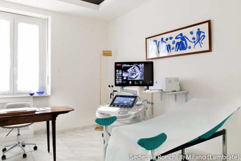
During an obstetric ultrasound, the patient lies on the couch and sees the images that appear on the monitor. The doctor slides the ultrasound probe over the patient's abdomen after applying a special gel. In the room, the light is dimmed to allow optimal viewing of the images on the monitor. The success of ultrasound scans depends on the position assumed by the fetus inside the uterus, the thickness of the abdominal fat (the presence of excess abdominal fat could be a limiting factor in the ultrasound examination). Obstetric ultrasound has no standard duration: the examination may be longer or shorter, depending on the clinical case.
Obstetric ultrasound preparation standards
It is not necessary to be fasting to perform an obstetric ultrasound. In the two days prior to performing an obstetric ultrasound, the application of creams and ointments on the belly should be avoided, as these may make ultrasound viewing more difficult.
Book an ultrasound
OUR OFFICES
CHOOSE WHERE TO PERFORM THE EXAMINATION
MILAN:
- Studio Ginecologico Milano, in Via Ronchi 8, mezzanine floor, in front of train station exit and metro Lambrate, Side via Rombon. Access for people with disabilities. MELEGNANO:
- Medical office located downtown at 63 Castellini Street, staircase f, 9th floor, 3 elevators present, access for people with disabilities.


