SONOHYSTEROSALPINGOGRAPHY (SONOHYSTEROGRAPHY) IN MILAN
SUPPORTING ULTRASOUND, ACCURATELY DESCRIBES ANY PATHOLOGY OF THE UTERINE CAVITY

WHAT IT'S FOR
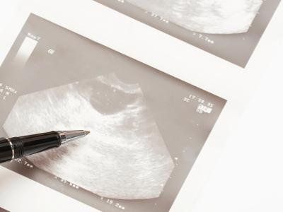
Sonohysterosalpingography (Sonohysterography) is an examination that complements vaginal ultrasound, performed with a two-dimensional (2D) or three-dimensional (3D) probe. It accurately describes internal pathologies of the uterine cavity projecting within it, already suspected by vaginal ultrasound. Diagnoses endocavitary neoformations: polyps, fibroids, and uterine synechiae that are the cause of abnormal uterine bleeding, menorrhagia, metrorrhagia, or amenorrhea and infertility. Assesses tubal patency, an essential condition for spontaneous conception. Finally, it excludes uterine malformations. It raises the suspicion of cervicohisthmatic incontinence of the cervical canal, a cause of polyabortion and premature delivery in the second trimester of pregnancy. Its reliability is very high in terms of diagnosis. It has, in fact, drastically reduced the use of hysteroscopy and hysterosalpingography (in which X-rays are used).
HOW IT IS PERFOPRMED
It requires no previous preparation. It is performed in a properly equipped outpatient clinic. It is quickly performed. Its average duration is 15-20 minutes. It is well tolerated. The woman may experience some transient discomfort during its performance. Side effects (pelvic menstrual algias, vaginal bleeding, vaso-vagal reaction) are rare, mild and short-lived. However, it may be good prudential practice for the patient to present accompanied by a person who can take her to her home.
The technique is very simple. The patient positions herself on the gynecological couch with her legs spread apart and resting on the side supports. Introducing the speculum into the vagina, the gynecologist inserts a thin, sterile, latex catheter inside the uterus. Then, gently removing the speculum, he inserts the transvaginal probe into the vagina and injects contrast medium inside the uterine cavity through the thin catheter. This contrast medium dilates the walls of the uterine cavity and makes the internal cavity of the uterus visible ultrasound, the exact location of any neoformations within the uterine cavity, and tubal patency.
Book an examination
OUR OFFICES
CHOOSE WHERE TO UNDERGO SCREENING
MILAN:
- Studio Ginecologico Milano, in Via Ronchi 8, mezzanine floor, in front of train station exit and metro Lambrate, Side via Rombon. Access for people with disabilities
MELEGNANO:
- Medical office located in Via Castellini 63, staircase f, 9 floor, 3 elevators present, access for people with disabilities


