


THE ULTRASOUND EXAMINATION IN MILAN
AN IMPORTANT INVESTIGATIVE TOOL WITH LOW COSTS
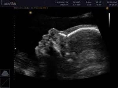
Exploits the properties of ultrasound (high-frequency sound waves) to collect images of internal organs and structures in the lower abdominal area, including the uterus, vagina, fallopian tubes, cervix, ovaries, and bladder.
WHAT IT'S FOR
Gynecologic (pelvic) ultrasound is a useful diagnostic tool associated with gynecologic examination. It is indicated for all persons for whom it is necessary to draw information (such as size, shape, position, appearance, or thickness) regarding the affected organs of the female genital system.
Gynecologic ultrasonography aims to study the morphology of all organs in the lower abdominal area (bladder, uterus, and ovaries) to reveal any atypical solid masses (fibroids or tumors) or cystic formations, effusions, foreign bodies, or structural malformations.
It allows the pelvic organs (uterus, tubes, ovaries, bladder) to be visualized and to detect the possible presence of pathologies such as: endometrial thickening, endometrial polyps, uterine myomas (fibroids, uterine leiomyomas), ovarian cysts, tubal abnormalities (sactosalpinges) and other pathologies.
The main function of gynecologic ultrasonography is to detect abnormal masses of the female genital system. The such finding makes possible a timely diagnosis that can follow up with effective therapy in counteracting any pathology observed during the examination.
In women, in particular, the examination makes it possible to investigate the causes of the following manifestations or conditions:
- Infertility or abnormal vaginal bleeding;
- pelvic pain;
- amenorrhea;
- benign and malignant tumors of the ovaries and uterus;
- presence of fluid or masses in the endometrium or myometrium (uterine muscle tissue);
- changes in the fallopian tubes;
- location of an intrauterine contraceptive device (IUD, IUD).
how it is performed
Free of contraindications, it can be performed by two techniques:
TRANSADDOMINAL, placing a probe on the woman's abdominopelvic wall (i.e., belly). The suprapubic (external) procedure involves the doctor sliding the ultrasound probe directly over the skin after applying a thick, transparent gel. It should be performed when the bladder is full to allow a more accurate assessment of the clinical picture. It is important not to urinate but to drink about half a liter of water at least one to two hours before the gynecological ultrasound. This is the only ultrasound modality in the virgin patient.
TRANSVAGINAL involves the introduction of the probe into the vagina. The procedure causes little discomfort, similar to that of a gynecological examination, has greater diagnostic accuracy, being inserted into the space between the cervix of the uterus and the vagina, in contact with the internal organs.
The physician who prescribes or performs the examination suggests the most appropriate approach for each patient.
Book an ultrasound
The sites
Choose where to undergo the examination
MILAN:
- Studio Ginecologico Milano, in Via Ronchi 8, mezzanine floor, opposite train station exit and metro Lambrate, Lato via Rombon. Access for people with disabilities.
MELEGNANO:
- Medical practice located downtown at Via Castellini 63, staircase f, 9th floor, 3 elevators present. Access for people with disabilities.


