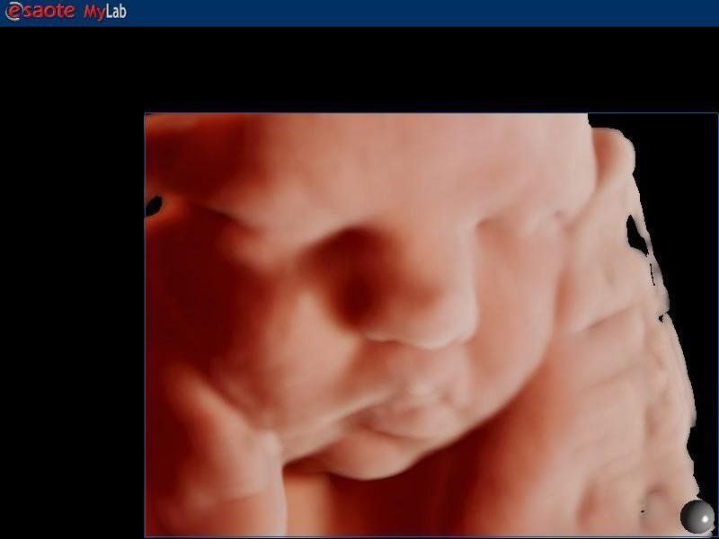
3D AND 4D THREE-DIMENSIONAL OBSTETRIC ULTRASOUND
WHAT IS IT FOR?
Obstetric (3 e) 4 D ultrasound provides moving, therefore "more realistic" three-dimensional images of one's baby. It allows for the first time to observe life in a woman's womb. It is like seeing one's child "live". Visual reconstruction gives parents an idea of what the fetus looks like in reality. It is exciting to see for the first time the baby moving, smiling, grimacing or sucking its finger. Fetal movements can be visualized: from gross limb movements to facial expressions (smiling, yawning, sucking, etc.). Sometimes the moving image is so timely that it shows parents their baby moving, even observing facial expressions so that they can begin an emotional and relational relationship even before birth. It produces a great emotional impact in parents. The mother-baby relationship (also called "bonding") seems to be positively affected by ultrasound. It helps to strengthen the emotional bond already present between mother and unborn child. In some cases it has such a positive influence that it even prompts the mother to improve her lifestyle such as quitting smoking or drinking alcohol. In this process, three-dimensional ultrasound seems to have a greater capacity than traditional ultrasound in increasing the bond between the mother and her baby. For' the examination to be clear and the fetal face clearly visible, it is important that the fetus is properly positioned and that there is a good stratum of amniotic fluid between the fetus and the probe and that the patient's echogenicity is good.
Three-dimensional ultrasound uses ultrasound in the same way as traditional ultrasound, so it has no risk and no particular contraindications to its use. The term 4D was introduced to define three-dimensional ultrasound in real time, in fact the fourth dimension is time.
In special cases, its field of application is prenatal diagnosis. It identifies the exact position of the gestational sac within the uterus in doubtful cases. It can be useful in the study of fetal anatomy, in the diagnosis of any abnormalities of internal organs, the spine, the four limbs, the surface anatomical structures of the fetal body, and finally the umbilical cord. It improves the definition of defects of the facial massif (particularly the lip and palate). Identifies the location of a neural tube defect, present on the spine. It more accurately defines limb malformations and abdominal wall defects (such as gastroschisis and omphalocele). However, the main limitations of this method are fetal movements and the small amount of amniotic fluid near the anatomical structures being studied.
WHEN TO PERFORM IT?
Obstetric 3 and 4 D ultrasound can be performed at any time during pregnancy. The best time is from 24 weeks to 28-29 weeks of gestation. At that gestational age the image of the fetus is sharper. Although already in the first trimester it is possible to perform the 3-D and 4-D examination realizing how already at 12 weeks the fetus is fully formed and in full activity.
METHODICS
Three-dimensional ultrasound is based on computer reconstruction and processing of normal two-dimensional ultrasound images. Three-dimensional (3D) images can be obtained, i.e., still images that are analyzed from various viewpoints and in three dimensions, or four-dimensional (4D) images, where the fourth dimension is time, i.e., you have three-dimensional images in motion and in real time.

Book a 3D or 4D echo
OUR OFFICES
CHOOSE WHERE TO TAKE THE EXAM
MILAN:
- Studio Ginecologico Milano, in Via Ronchi 8, mezzanine floor, in front of train station exit and metro Lambrate, Side via Rombon. Access for people with disabilities. MELEGNANO:
- Medical office located downtown at 63 Castellini Street, staircase f, 9th floor, 3 elevators present, access for people with disabilities.


